We use cookies to make your experience better. To comply with the new e-Privacy directive, we need to ask for your consent to set the cookies. Learn more.
What is Torn Knee Meniscus?
The lateral and medial menisci are crescent-shaped fibrocartilaginous structures that collectively cover approximately 70% of the articular surface of the tibial plateau. The primary function of the menisci is load transmission and shock absorption through the tibiofemoral joint. 70 percent of each meniscus is made up of a network of type1 collagen which is arranged primarily in circumferential direction.
Meniscal tears are categorized by both their shape and location when visualised on MRI.
Knee meniscal injuries are common with an incidence of 61 cases per 100,000 persons and a prevalence of 12% to 14%. There is an increased incidence of meniscal tears with anterior cruciate ligament (ACL) injury ranging from 22% to 86%.
Causes
The meniscal tears occur due to rotational or shearing forces placed across the tibiofemoral joint, especially in situations in which increased axial load is placed through the menisci. Trauma can also lead to meniscal tear or tears occurring simultaneously with bony lesions and damage to the primary stabilising ligaments of the knee like Anterior cruciate ligament (ACL) and Medial cruciate ligament (MCL).
Sign and symptoms
Clinical presentation of the patient with a meniscal tear is variable depending on the mechanism of injury and degree of concomitant tibiofemoral insults. Patient presents with the sensation of a "pop," with immediate effusion of the knee during high-impact activity or trauma is associated with an ACL tear which might be associated medial meniscal tear, whereas effusion that develops more gradually over the course of 24 hours is more indicative of an isolated meniscal tear.
The patient also complains of pain over the anteromedial or anterolateral joint line. On physical examination, the knee must be looked for oedema. The joint line must be palpated. Also, standing and supine range of motion (ROM), muscle strength testing etc. must be performed.
Diagnosis
In case of meniscal tear suspicion, radiographs must be recommended that include AP, lateral, oblique, sunrise and weight bearing views for the assessment of any underlying bone pathologies. MRI imaging is the best mode of imaging to diagnose and characterize meniscal tears.
General management
The management of acute painful, oedematous meniscal tears must include RICE (Rest, Ice, Compression and Elevation). Medications for symptomatic relief must be recommended. For simple tears, it is reasonable to perform a 4צ6-week course of relative rest and physical therapy to determine if spontaneous healing and return to the desired level of function will occur.
Warning: Above information provided is an overview of the disease, we strongly recommend a doctor's consultation to prevent further advancement of disease and/or development of complications.
Disclaimer: The information provided herein on request, is not to be taken as a replacement for medical advice or diagnosis or treatment of any medical condition. DO NOT SELF MEDICATE. PLEASE CONSULT YOUR PHYSICIAN FOR PROPER DIAGNOSIS AND PRESCRIPTION.
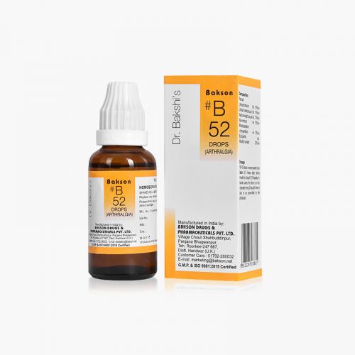
- #B 52 DROPSSpecial Price ₹ 160.00 Regular Price ₹ 200.00
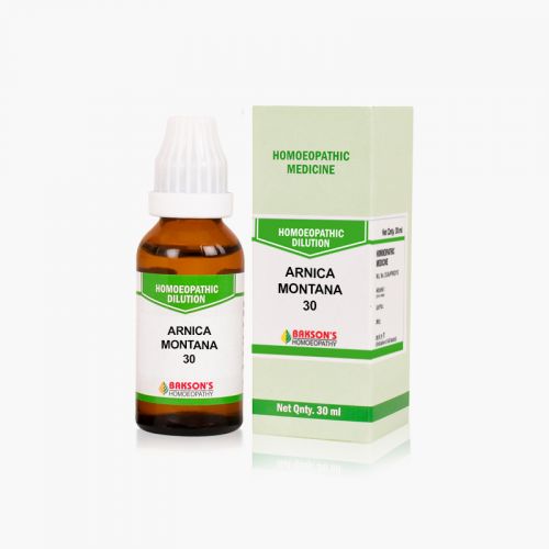
- ARNICA MONTANA 30₹ 100.00
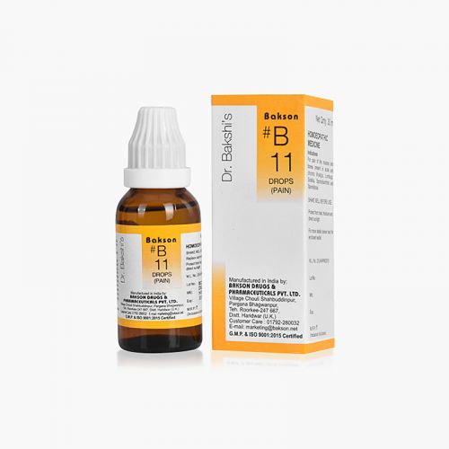
- BAKSON #B 11 DROPSSpecial Price ₹ 160.00 Regular Price ₹ 200.00
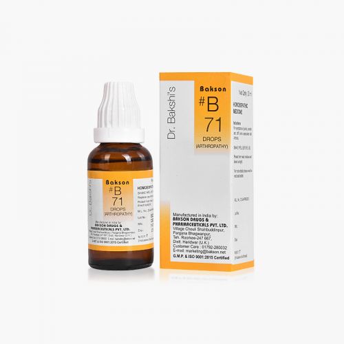
- BAKSON #B 71 DROPSSpecial Price ₹ 160.00 Regular Price ₹ 200.00
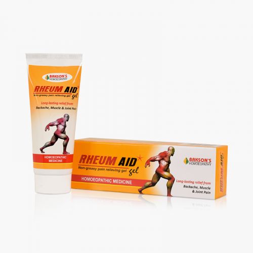
- BAKSON RHEUM AID GELSpecial Price ₹ 84.00 Regular Price ₹ 105.00
-
- BCT # 19 (JOINT & MUSCLE PAIN)-250TABSpecial Price ₹ 84.00 Regular Price ₹ 105.00
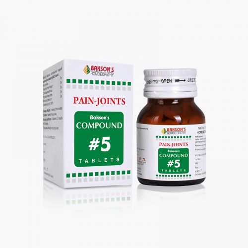
- COMPOUND #5 TABLETS-100TABSSpecial Price ₹ 108.00 Regular Price ₹ 135.00
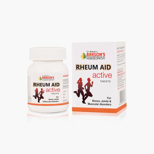
- RHEUM AID ACTIVE TABLETS - 75 TABSSpecial Price ₹ 172.00 Regular Price ₹ 215.00
-
-
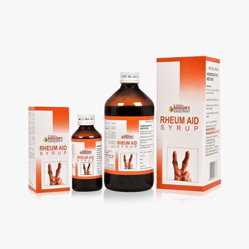
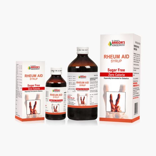
- RHEUM AID SYRUP (SUGAR FREE)As low as ₹ 112.00
